animal cell electron microscope
You see that many features are in common. Electron microscopy is in addition a valuable tool for the investigation of the most subtle effects of disease-causing organisms and toxic substances on animal cells and tissues.

Visualization Of The Cell Using Em Scanning Electron Microscope Microscopic Photography Cell
Monday April 5th 2021.

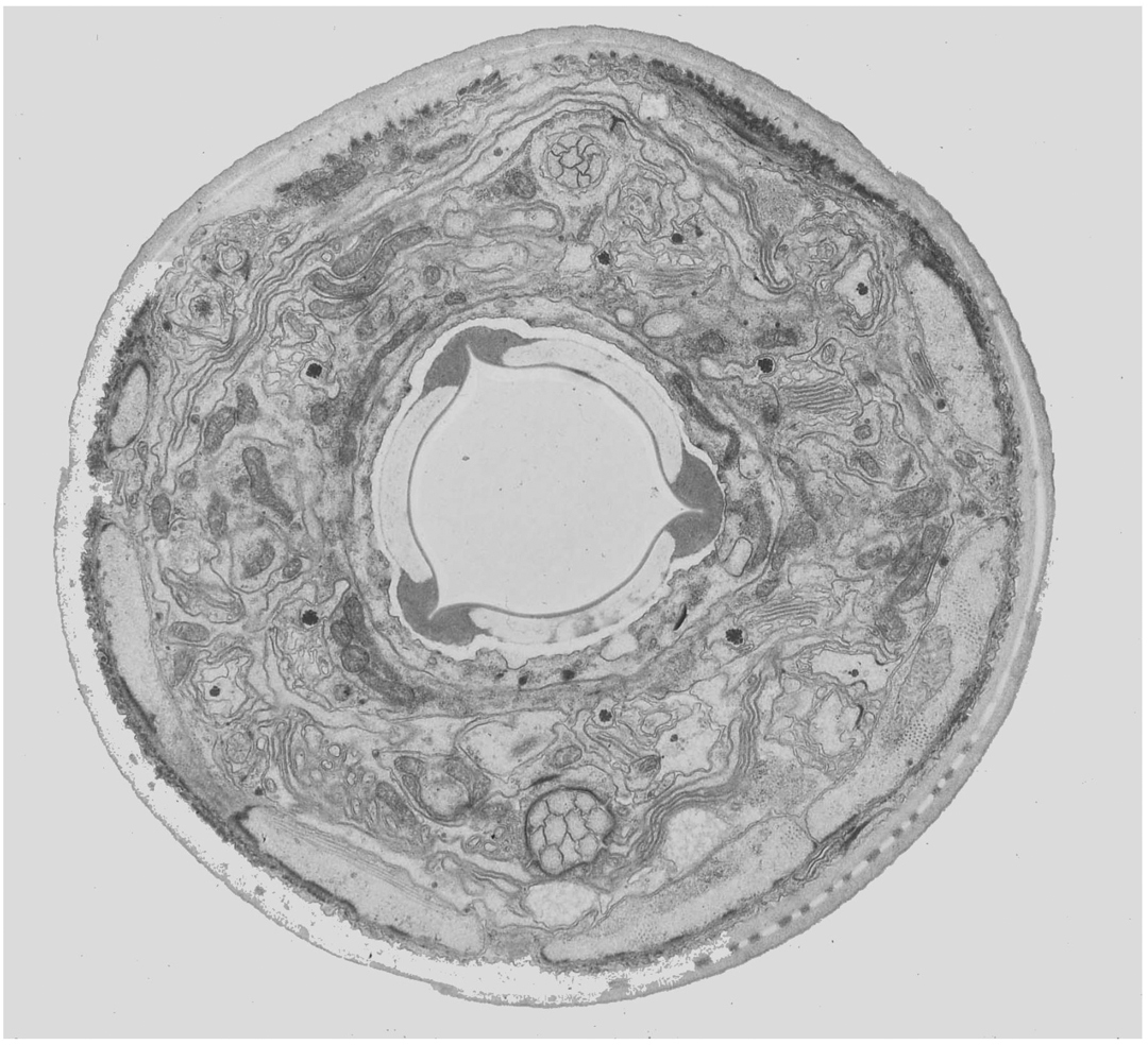
. Here is an electron micrograph of an animal cell with the labels superimposed. Three-dimensional views of the surfaces of cells and tissues are obtained by scanning electron microscopy. 1 The name mitochondria was given by.
Get started in a snap. A typical animal cell as seen in an electron microscope Medical Images For PowerPoint 1. Animal cells are typical of the eukaryotic cell type enclosed by a plasma membrane and containing a membrane-bound nucleus and organelles.
Diagram Of Animal Cell Under Electron Microscope Labeled. The electron microscope is. Ad Digital microscopy with automated brightfield fluorescence and Digital Confocal imaging.
A typical animal cell as seen in an electron microscope Medical Images For PowerPoint 1. Ad Digital microscopy with automated brightfield fluorescence and Digital Confocal imaging. Get started in a snap.
Explore the structure of. Within the cell there is a shape of round with a circular structure of granulated part on the. Do more than cell counting.
Do more than cell counting. Animal cell under electron microscope. Diagram Of Animal Cell Under Electron Microscope.
Observing a wide range of biological processes and animal cell under light microscope is easier due to advances in microscopic techniques. The transmission electron microscope TEM is used to examine thin slices or sections of cells or tissues the scanning electron microscope SEM has a large depth of field so can be used to. Thats the major difference between plant and animal cells under microscope.
It is an electron micrograph of cells largest and most important organelle the mitochondria and is characterized by the following features Fig. Typical Animal Cell Pinocytotic vesicle Lysosome Golgi vesicles Golgi vesicles. Generalized Structure of a Plant Cell Diagram.
The transmission electron microscope TEM is used to examine thin slices or sections of cells or tissues. Transmission electron microscope TEM and Scanning electron microscope SEM. We have got 7 pic about diagram of animal cell seen under electron microscope images photos pictures backgrounds and more.
However only TEMs can see inside a cell. To use a light microscope to examine animal or plant cells diagram. The diagram below shows the general structure of an animal cell as seen under an electron microscope.
560 X 364 Pixel Electron Microscope Image Animal Cell And Organelles Labeled Animal Cell Plasma Membrane Organelles. A typical animal cell as seen in an electron microscope Medical Images For PowerPoint 1. You can observe this epithelial animal cell under microscope with high power.
There is two type of electron microscope. When storing use a plastic. The shapes of isolated macromolecules that have been shadowed with a heavy.
The animal cell is more fluid or elastic or malleable in structure. As you can see in the above labeled plant cell diagram under light microscope there are 13 parts namely Cell membrane. So lets begin by drawing a rough-oval.
TEMs have a maximum magnification of around 1000000 but images can be.

Animal Cell Organelles Cell Organelles Organelles

Animal Cell Structure Cell Organelles Electron Microscope

Visita Il Nostro Sito Templedusavoir Org Electron Microscope Electron Microscope Images Microscopic Photography

Animal Cell Organelles Sauna Design

Electron Micrograph Of Me Celula Animal Biologia Microscopio Electronico

Gulf War Illness Not In Veterans Heads But In Their Mitochondria Scienceblog Com Mitochondria Animal Cell Organelles Animal Cell

Cell Organelles Animal Cell Organelles

Image Result For Diagram Of Plant And Animal Cell Under Electron Microscope Celula Animal Ciencias Verdades Absolutas
Cells Under Electron Microscope Google Search Animal Cell Structure Cell Diagram Animal Cell

Wellcome Education On Twitter Microscopic Images Electron Microscope Images Organelles
Cell Biology Biology Animal Cell

Pin On Cell

560 X 364 Pixel Electron Microscope Image Animal Cell And Organelles Labeled Animal Cell Plasma Membrane Organelles

Animal Cell Structure And Organelles With Their Functions Jotscroll Organelles Animal Cell Cell Diagram

Golgi Apparatus Tem Cell Organelles Animal Cell Macro And Micro

Electron Microscopy Cell With Organelles Biology Art Electron Microscope Images Microscopy

Muppets Animal Drawing At Paintingvalley Com Explore Collection Of Muppets Animal Drawing Cell Diagram Animal Cell Structure Animal Cell

Pin On Ib Biology Hl

An Electron Micrograph Of A Mouse Liver Cell Dna Learning Center Electrons Cell Learning Centers